
Why a congenital defect could cause calf losses
 © Adobe Stock
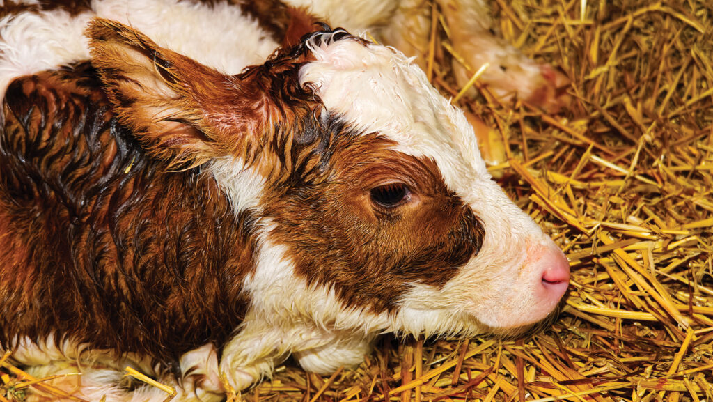
 Ask a dairy or beef farmer if they have ever had a calf born with atresia, a condition commonly known as “waterbelly”, and the chances are they will say no.
But Prof John Mee, a principal veterinary research officer at the Irish farm research body Teagasc, says atresia is likely to be more common than farmers believe.
Cases are not often picked up because so few calves are presented at disease surveillance centres for post-mortem, he points out.
See also: How two herds have reduced incidence of stillbirth
“In general, the submission rate of calves that die of all kinds of conditions is extremely low in comparison to the number that die on farms,’’ says John, who has an interest in congenital defects in calves.
He had previously been involved in a surveillance study of congenital defects, collecting dead calves from farms for examination.
“In doing this we discovered that congenital defects were a much more common cause of death on farms than we previously thought,’’ he says.
“And while we thought skeletal or cardiac defects would be the most common [ones], it turned out that the most common was atresia.’’
Symptoms
Intestinal atresia results in complete blockage of the bowel in the foetal calf during the early stage of pregnancy.
It is sometimes called waterbelly because the blockage causes intestinal fluid to build up during pregnancy, so the calf may be born with a stomach that is full of fluid and swollen.
A study, carried out over an eight-year period and involving 40 Irish dairy herds and 56,454 calves, looked at the role genetics plays in atresia.
In total, 197 cases of intestinal atresia were examined at the post-mortem laboratory in Moorepark Research and Innovation Centre.
Here, samples were collected for genetic analysis, with associated risk factor information taken both from the herds of origin and the affected calves.
A case-control, genome-wide association study was also done to detect genomic variants associated with intestinal atresia.
The study found that the overall incidence of intestinal atresia was 0.35%, with some herds recording up to 1.34%.
It revealed that intestinal blockage was twice as common in male calves than in females, and in the progeny of older cows (calves born to dams that had calved more than three times).
Clusters occurred in some herds – the breed with the highest incidence was the Jersey, at 1.02%, compared with Holstein-Friesian at 0.07%.
There was greater incidence among the progeny of three related Jersey sires, which the researchers say suggests a gene for intestinal atresia was segregating within this family.
There was no evidence of a sire-effect among the progeny of Holstein-Friesian sires.
John says a reason for the higher incidence in Jerseys could be that fewer are used for breeding than Holstein-Friesians.
A genetic link was also detected in a particular Jersey family, rather than in Jerseys in general, he adds.
The signs of atresia
With atresia, blockage can occur in different parts of the bowel or, less commonly, the anus.
In a Teagasc study in Ireland, 83% of calves were affected in the small intestine, 14% in the large intestine and, for 3%, the anus.
While an affected calf may appear normal or swollen at birth, it may take a few days for someone to notice that it is not passing dung.
Initially a calf may drink normally, but will then stop drinking, lie down a lot and become more swollen.
This is exacerbated if more milk is fed by tube when a calf is not sucking. Without surgery, a calf usually dies within seven days of birth, or is euthanised.
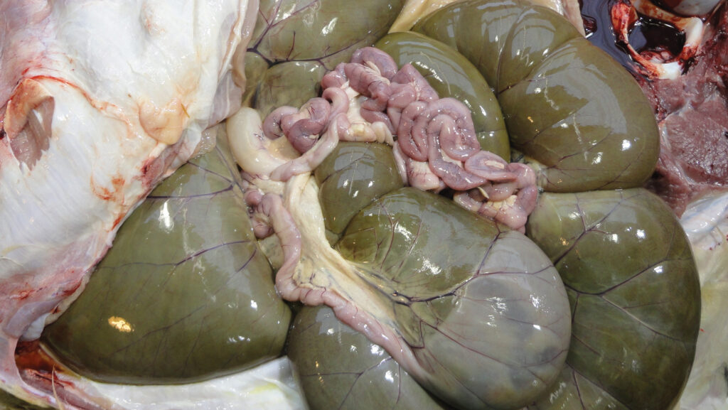
© John Mee
Another indicator of the condition is when a newborn calf has no meconium coming from its back end.
“Farmers with a history of these cases should examine the anus of the calf immediately after birth and if they can’t see any meconium they should put a finger in the anus to see if they can digitally express some faeces,’’ says Teagasc’s Prof John Mee.
Although surgery can be done, he suggests the success rate is poor as animals do not have productive lives afterwards.
“Now that we have established a genetic link, there is another contra argument against surgery because if this is a genetic and inherited [condition], you will be propagating atresia down to the next generation.’’
Inbreeding effect
“Relatively speaking, the likelihood of inbreeding or co-ancestry would be higher in a Jersey because there are fewer to choose from, but we didn’t examine that in this study so we can’t be sure of it, though it is a likely reason,’’ John says.
“We did find Jersey sires that had a direct genetic link to this condition and we couldn’t find it in Holsteins, even though we had far more animals to examine than Jerseys.’’
The study results provide further understanding into the causes of atresia, helping to inform decision-making on farms. If a farm has had cases in the past, John recommends the farmer has a discussion with their semen supplier.
“Ask the supplier if they have been doing any recording on this, and do they know of any risks in the sires they are presenting for breeding.’’
John is aware of rare instances in herds where a cow has produced successive calves with atresia.
“In cases like this, it is worth looking back at herd records to see if that animal is the heifer of a cow that also had cases, to look for the familial lines in the female side independent of the sire family.
“It won’t delete many cases but, if you have a high number, you need to look at both sides.’’
© Adobe Stock
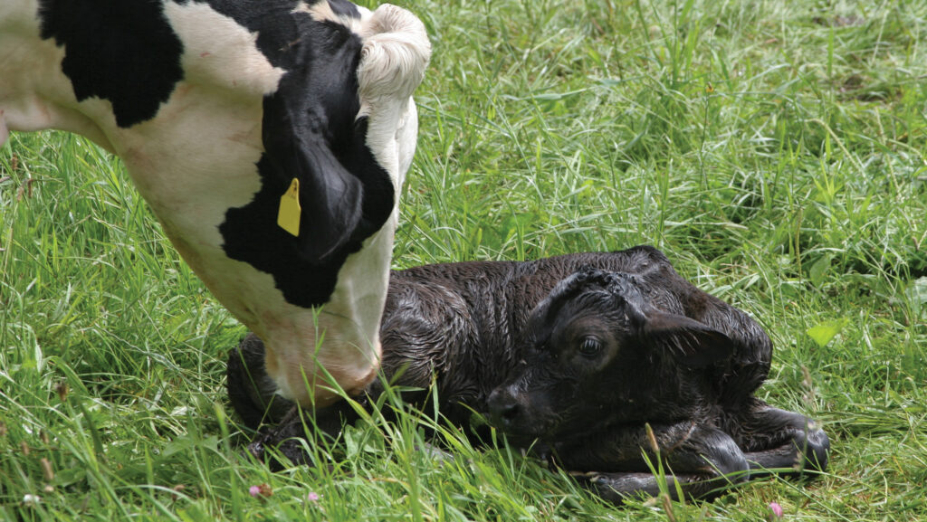
Calf losses
Any farm that experiences calf losses in the first 10 days after birth should question if atresia is a cause in these mortalities.
Aside from genetics, John points out that a risk factor for atresia is manually examining a cow to establish if she is in calf, a technique usually used to detect pregnancy as early as six weeks.
In the early stages of development, the bowel is in the umbilical cord – outside the foetus – and it can get damaged when a foetus is examined manually in those first six weeks of pregnancy.
“This hasn’t been proven, it is a hypothesis, but on a farm with an ongoing problem with atresia, there is good reason for the farm to discontinue that intervention,’’ he says.
“If they want to continue pregnancy diagnosis for practical management reasons, they should switch to ultrasound scanning.’’
He points out that there is no known relationship between mineral or nutritional deficiencies and atresia.
The UK has no surveillance data on congenital defects, but bovine veterinary specialist Owen Atkinson has encountered atresia in both calves and lambs.
He has seen cases where the animal has a combination of a distended bowel and colic, and is unable to pass faeces.
Identify at post-mortem
Usually the animal has needed to be euthanised, but Owen has successfully performed surgery on a beef calf that the owner did not intend to use for breeding, creating a fistula for the calf to excrete through.
However, the majority of cases he has seen have been at post-mortem, after the condition had gone unnoticed by the farmer and the calf had died.
He says the farmer would have been unaware of the congenital defect because, in many cases, the calf appears bright and normal at birth.
Manual palpation to detect pregnancy has largely been replaced by ultrasound scanning, he adds.
“With ultrasound, the evidence is that the risk of causing damage to the foetus, in skilled hands, is minimal.’’